The conditions used for microscopy are often not “physiological” conditions. If we are talking about live imaging, then the cells are usually in culture, placed on a glass surface and grown in an artificial media. In many cases, we use genetically encoded fluorescent markers, that are rarely inert. These are acceptable and known limitations of the system.
However, when we think about microscopy, we do not often consider to effect of the light itself. The light we use can have deleterious effect. For instance, UV light is known to cause damage to cells, e.g. creating reactive oxygen species (ROS), creating thymidine dimers, cross-linking macromolecules and probably more. That is why people tend to limit the use of fluorescent proteins that are excited by UV light (such as blue fluorescent proteins or some photoactivated proteins).
Yet, visible light is considered relatively harmless. Now, a new study suggests that visible light, particularly blue-green light (which is used to excite green-yellow fluorescent proteins), can affect the metabolic state of the cell. How?
Well, all cells contain light absorbing molecules. Some molecules function as light sensors or light energy harvesting molecules: Chlorophyll, cryptochromes, phytochromes, photoreceptors, rhodopsins, and fluorescent proteins. Other molecules absorb light, e.g. pigments.
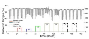
However, some cells do not have obvious light sensitive molecules. For example, the budding yeast. In this paper, Carl H. Johnson’s lab looked at the effect of visible light on yeast respiratory oscillations (YRO) and oxidative stress. Apparently, under some conditions, yeast show 1-6hr oscillations of oxygen consumption and metabolite productions. surprisingly, shining white, blue or green light (but not red) has shortened the cycle and decreased the amplitude of oxygen consumption. There was also increased expression of antioxidant enzymes, and increased light sensitivity to oxidative-stress mutants.

The effects of different spectra of light on the YRO. Oscillations were initiated in the dark until stable oscillations formed (black line, left y-axis). Then 12-h treatments of red, blue, or green light were administered (colored lines matching color of light, right y-axis) with 12 h of darkness between treatments. After the application of colored light, two 12-h white light treatments were given. Light intensities of each treatment are shown on the right y-axis and are indicated by numbers under each of the colored or gray lines showing light treatment. Source: Robertson J B et al. PNAS 2013;110:21130-21135
Based on several other assays, the authors suggests the blue-green light harms cytochromes in the respiratory electron transport process. Photoinhibition of the electron transport causes accumulation of ROS and oxidative stress response. It is hypothesized that the shortened oscillation periods and reduced amplitude of oxygen consumption (= less respiration) is a cellular mechanism to reduce the photoinhibition to electron transport, thus reduce oxidative damage.
What does this mean to microscopists?
It only suggests that long term exposure to light (either in prolonged live imaging, or just the incubation conditions) can affect the metabolic state of the cell and therefor may have either deleterious effect on the health of the cell, or create false positive or false negative results, particularly in experiments that are designed to study respiration and oxidative stress. Obviously, if you have light sensors or light harvesting molecules, their absorbance wavelengths need to be taken into account.
Practically, it is always a good advice to minimize the exposure to light (which we do anyway to reduce photobleaching).
![]() Robertson JB, Davis CR, & Johnson CH (2013). Visible light alters yeast metabolic rhythms by inhibiting respiration. Proceedings of the National Academy of Sciences of the United States of America, 110 (52), 21130-5 PMID: 24297928
Robertson JB, Davis CR, & Johnson CH (2013). Visible light alters yeast metabolic rhythms by inhibiting respiration. Proceedings of the National Academy of Sciences of the United States of America, 110 (52), 21130-5 PMID: 24297928


