In eukaryotes, the DNA is packages tightly in nucleosomes, which are composed primarily out of histone proteins. There are four major types of histones (1,2,3 & 4). Extensive work has been done on how histones facilitate and regulate transcription. It turns out that there are multiple post-translational modifications on histones, such as methylation and acetylation that are linked to transcription regulation. The majority of the studies use a method called chromatin immunoprecipitation (ChIP) to study these modifications. In essence, an antibody specific for a certain modification is used to affinity-purify only modified histones, along with any DNA region they are associated with. Thus, one could get a map of the specific modified histone along the chromosomes and correlate this locations with transcription activity, ChIP maps of other transcription related proteins etc…
There are two problems with this approach. The first, since the cells are fixed, the time resolution is limited to several minutes, at best. Second, the results are an average of the entire cell population, and therefore factors considered linked may not actually be present in the same cell, same genomic location at the same time.
So, Timothy Stasevich et al. tried a different approach by using a novel method to image histone modifications in live cells.
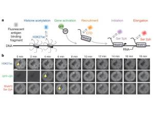
This method is called Fab-based live endogenous modification labelling (FabLEM). Basically, these are the antigen binding fragments (Fab) of a specific monoclonal antibody, conjugated to a fluorescent dye. The Fabs are small enough to enter the cell and the nucleus.
By using this system, they reached a temporal resolution of about 10 seconds!
They used a cell line that harbours a 200-copy gene array (i.e. an array of 200 copies of the same gene) whose promoter is controlled by a GFP-labeled glucocorticoid receptor (GR). Why this cell line? This gene array manufactures a very concentrated fluorescent signal, composed of 200 molecules of whatever dye associates with it, in a very small volume. This allows for a good signal to noise ratio compared to the background of the free or chromatin associated fluorescent-labeled molecules.
For the first experiment, they looked at acetylation on lysine position 27 of histone H3 (H3K27ac), in regard to GR-GFP accumulation (transcription initiation) and the state of RNA polymerase II (RNAP2). The C-terminal domain (CTD) of RNAP2 can be unphosphorylated, or phosphorylated on serine 5 (marking transcription initiation) or serine 2 (marking transcription elongation) of the ~50 heptad repeats that it contains.

a, Illustration of the FabLEM strategy. b, Examples from a FabLEM experiment showing that the gene array (yellow arrow) is acetylated at H3K27 before activation by GFP–GR and accumulation of elongating RNAP2 (Ser 2ph). Scale bar, 5 μm. Source: Stasevich et al (2014) Nature 516:272.
They initially found that the array is hyper-acetylated before induction, and the level of H3K27ac drops after induction. Here, also, comes the advantage of single-cell experiments. They found variability between cells before induction by the hormone – some cells with high H3K27ac and some with low level. So they checked whether these two populations differ in their transcription kinetics. They found no effect on Ser5 phosphorylation, but significant higher levels of GR-GFP recruitment and Ser2 phosphorylation in the high H3K27ac group. This indicated that H3K27ac levels can affect transcription factor recruitment as well as transcription elongation.
To examine if there is a causal connection between K3K27 acetylation and transcription elongation, the researchers perturbed the acetylation (2 experiments by 2 different approaches) and again found that though initiation (Ser5-P) was unaffected, Ser2-P in cells with reduced H3K27ac was also diminished. Consistent with this notion, they found that the protein AFF4, a key component of the elongation complex, which was shown to interact with the H3K27 acetylase p300, is recruited less efficiently to arrays with low H3K27ac.
To conclude: specific protein modifications has been difficult to image in live cells due to the technical issue of “how to specifically fluorescently tag them”. The use of FabLEMs can solve this problem. From discussing FabLEMs with Tim in the past, I understand that the Fabs are constantly binding on and off every few seconds, so that there is a (small) time window for the endogenous regulating protein which is supposed to bind the antigen to actually bind. But, we should take into account that the FabLEMs probably do perturb the biological function of any specific single protein they bind to. But, in these experiments, with an array of 200 genes, and hundreds or more of H3 proteins on each gene, this effect is limited. Similarly, the CTD of each pol II contains ~50 heptad repeats. though it is not clear how many repeats are phosphorylated when phosphorylation occurs, it is assumed that most, if not all, are phosphorylated. So in this case too its a game of numbers of the FabLEMS vs the regulatory proteins which bind these sites.
Still, these are issues that we need to consider when designing such experiments.
![]() Stasevich TJ, Hayashi-Takanaka Y, Sato Y, Maehara K, Ohkawa Y, Sakata-Sogawa K, Tokunaga M, Nagase T, Nozaki N, McNally JG, & Kimura H (2014). Regulation of RNA polymerase II activation by histone acetylation in single living cells. Nature, 516 (7530), 272-5 PMID: 25252976
Stasevich TJ, Hayashi-Takanaka Y, Sato Y, Maehara K, Ohkawa Y, Sakata-Sogawa K, Tokunaga M, Nagase T, Nozaki N, McNally JG, & Kimura H (2014). Regulation of RNA polymerase II activation by histone acetylation in single living cells. Nature, 516 (7530), 272-5 PMID: 25252976


“10 seconds”? That’s amazing!
LikeLike